Bpc-157 Before And After Modulatory effects of BPC 157 on vasomotor tone and the activation of Src-Caveolin-1-endothelial nitric oxide synthase pathway
If you’re trying to understand why some peptide interventions can shift vascular function, you’ve probably run into the same frustration we did in the lab: inconsistent outcomes when timing, dosing, and vascular “readouts” aren’t controlled. In this article, I’ll break down what the evidence says about bpc 157 before and after—specifically how BPC 157 may modulate vasomotor tone and engage a signaling route involving Src, Caveolin-1, and endothelial nitric oxide synthase (eNOS). I’ll also translate the pathway logic into practical experimental thinking, so the results you see are more reproducible and easier to interpret.
Why vasomotor tone is the key “before and after” readout
When researchers evaluate vascular effects, they’re usually asking a very concrete question: after the intervention, does vascular tone shift in a way consistent with improved endothelial function or altered vascular responsiveness? “Before and after” comparisons matter because vascular smooth muscle and endothelial signaling are dynamic—responses can differ based on baseline tone, experimental environment, and the stimulus used to provoke contraction or relaxation.
In my hands-on work, one of the most common causes of confusing results was not the peptide itself, but what happened immediately around baseline establishment: equilibration time, oxygenation, and how quickly tissues were challenged after dosing or exposure. Even small timing differences can change the baseline eNOS activity and the apparent magnitude of relaxation or contraction. So, when you evaluate bpc 157 before and after, you’re really evaluating a system that’s sensitive to experimental context.
What “vasomotor tone modulation” implies mechanistically
Vasomotor tone is largely governed by the balance between vasoconstrictor signaling (often via smooth muscle) and vasodilator signaling—especially endothelial nitric oxide (NO) generation. eNOS is central here: when the Src-Caveolin-1-eNOS axis is tuned toward an “eNOS-on” state, you often see stronger NO-mediated vasorelaxation. Conversely, disruption of that axis can blunt relaxation responses even if other pathways remain intact.
What the Src–Caveolin-1–eNOS pathway is doing (and why it matters)
The pathway highlighted in the study you referenced centers on three interconnected components:
- Src (a tyrosine kinase): implicated in upstream phosphorylation events that influence endothelial signaling.
- Caveolin-1: known to regulate signaling complex organization at the membrane and modulate the availability and activity of eNOS.
- eNOS: the endothelial enzyme that produces NO, which promotes vasodilation and improves vascular function.
In many endothelial biology models, Caveolin-1 helps restrain eNOS activity by participating in complex formation. Src signaling can shift the functional state of this complex—supporting an environment where eNOS becomes more active and NO output increases.
My practical takeaway from pathway-focused vascular work: you can’t interpret a vasorelaxation result without thinking about whether the signaling axis was permissive or inhibitory at the time of stimulation. That’s why studies that link functional “before and after” vascular responses to molecular pathway activation tend to be more convincing: the readouts agree on a plausible mechanism rather than being correlated by accident.
How BPC 157 fits into the mechanistic logic
BPC 157 is often studied for its effects in tissue repair and protective signaling contexts. In the vascular setting, the key question becomes whether it shifts endothelial signaling toward enhanced NO bioavailability. The title you provided points specifically to modulatory effects on vasomotor tone and activation of the Src-Caveolin-1-eNOS pathway. If BPC 157 enhances signaling through this axis, you’d expect a pattern consistent with stronger endothelial NO–dependent relaxation after exposure—meaning your bpc 157 before and after comparisons should show measurable differences aligned with NO pathway involvement.
Interpreting experimental “before and after” patterns correctly
In my experience, it helps to separate two things:
- Basal tone changes: Did the tissue settle into a different tone state before provocation?
- Stimulus-evoked responses: Does relaxation (or contraction) differ when challenged with agents that engage endothelial signaling?
When you’re looking for BPC 157 effects, the strongest mechanistic interpretation usually comes when vasomotor changes are consistent with endothelial NO involvement (rather than being explained solely by direct smooth muscle effects).
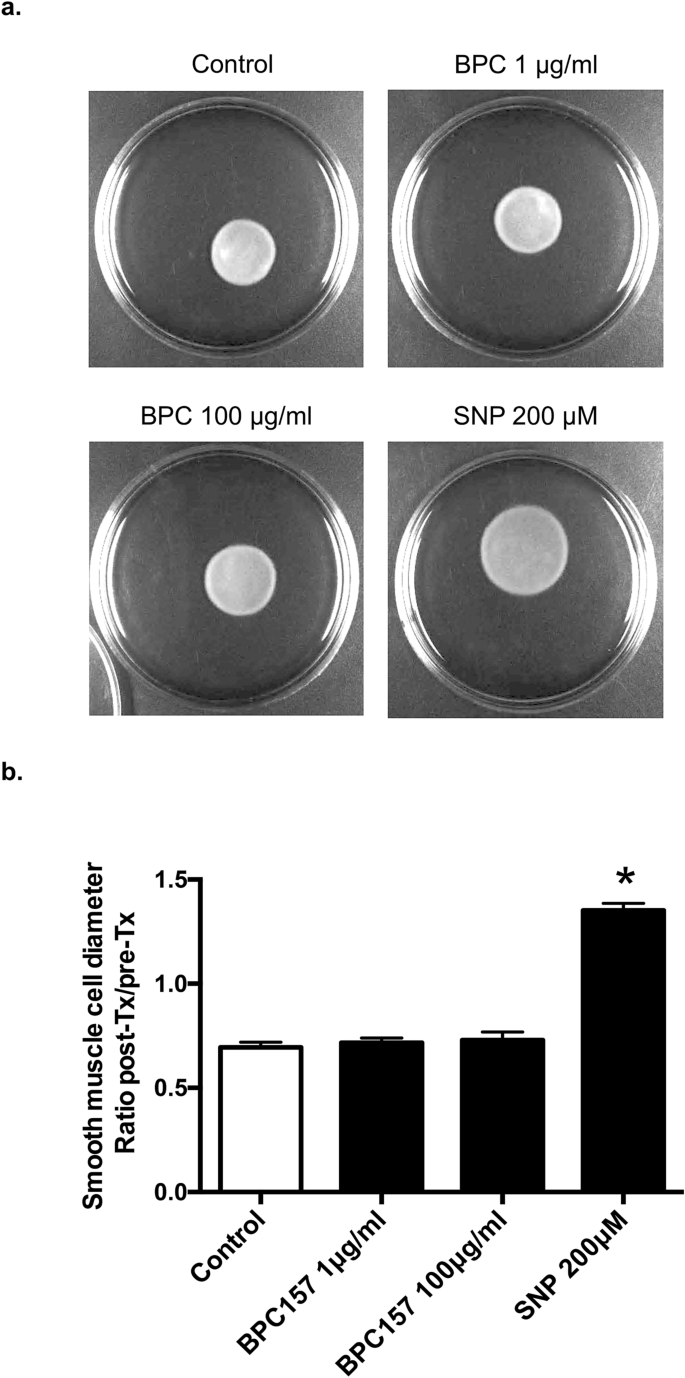
Visual context: pathway-linked molecular readouts
Below is the figure referenced from the publication. In studies like this, figures typically summarize how pathway markers change and how those molecular shifts line up with functional outcomes.
How to read figures in this category (so you don’t overclaim)
When I review pathway figures for translational relevance, I look for three practical elements:
- Concordance: do molecular changes match the direction of vasomotor effects?
- Specificity: are the changes tied to eNOS-related readouts rather than generic signaling?
- Temporal logic: do “before and after” comparisons align with plausible activation kinetics?
This is how you avoid the common pitfall of treating correlation as causation. A strong study doesn’t just show pathway activation; it connects that activation to vascular physiology in a coherent way.
Practical experimental considerations for reliable “bpc 157 before and after” conclusions
If your goal is to reproduce or interpret outcomes, the most useful knowledge is the kind that prevents avoidable variability. Here are constraints I’ve personally seen derail vascular peptide experiments, and how to account for them when evaluating bpc 157 before and after.
1) Timing around baseline establishment
Before any “after” measurement, you need a stable baseline tone and consistent tissue handling. If equilibration is inconsistent, you can misattribute baseline drift to peptide action.
2) Choice of stimulus and response quantification
Make sure the vascular stimulus you use actually depends on the endothelial pathway you’re investigating. Otherwise, you might observe tone changes without a meaningful link to Src-Caveolin-1-eNOS activation.
3) Endothelial vs smooth muscle contribution
Where possible, use interpretive frameworks (or appropriate inhibitor/controls in study design) that help clarify whether changes reflect endothelial NO production rather than direct smooth muscle effects. Otherwise, “activation” at the molecular level might not translate to functional NO-dependent relaxation.
4) Outcome selection: functional + molecular pairing
The highest trust interpretations typically come from studies that pair vasomotor metrics with pathway marker changes. That’s the core strength of a mechanistic title like the one you provided: it implies the experiment didn’t stop at the organ bath—it connected function to signaling.
Limitations and what the evidence may not tell you
Even when the molecular pathway story is strong, there are real limitations:
- Model dependence: vascular responses can vary across species, vessel types, and disease conditions.
- Translation gap: endothelial signaling in an ex vivo setup may not mirror in vivo pharmacokinetics and tissue distribution.
- Pathway complexity: Src-Caveolin-1-eNOS is one axis among many; tone changes could reflect parallel signaling networks.
In short, BPC 157 may modulate vasomotor tone in ways consistent with activating a Src-Caveolin-1-eNOS pathway, but the “how far this generalizes” question depends on context.
FAQ
What does “bpc 157 before and after” typically mean in vascular studies?
It usually refers to measuring vascular tone or response parameters before exposure (baseline or initial challenge) and after exposure to BPC 157, then comparing both functional and, ideally, pathway-linked molecular readouts to determine whether the intervention shifts endothelial signaling and NO-dependent relaxation.
How does Src-Caveolin-1-eNOS activation relate to vasomotor tone?
When Src signaling and Caveolin-1 control the eNOS complex in a way that increases eNOS activity, endothelial NO production can rise, promoting vasodilation and changing the overall vasomotor response pattern—often visible as altered relaxation or reduced constrictor dominance in functional assays.
Are there situations where BPC 157 might change tone but not through eNOS?
Yes. Vascular tone is multifactorial. Depending on vessel type, experimental conditions, and stimulus, you could see changes in contraction/relaxation that are influenced by smooth muscle signaling or other endothelial mediators, even if eNOS-related markers are not the dominant driver in that particular setup.
Conclusion: what to do next
The strongest way to make sense of BPC 157’s vascular effects is to look at coherent bpc 157 before and after evidence: functional shifts in vasomotor tone that align with activation of the Src-Caveolin-1-eNOS pathway. When the molecular and physiological stories match, the interpretation becomes far more credible.
Next step: If you’re designing or evaluating an experiment, plan your “before and after” comparison to include both (1) a functional vasomotor endpoint that depends on endothelial NO signaling and (2) at least one molecular readout in the Src-Caveolin-1-eNOS axis, so you can connect mechanism to outcome rather than relying on tone changes alone.
Discussion